Panretinal photocoagulation:
Panretinal photocoagulation (PRP) has long been a cornerstone in the management of retinal disorders, but alternative treatments are emerging as viable options for certain conditions.
What is Panretinal photocoagulation(PRP)?
PRP, a standard treatment for conditions like proliferative diabetic retinopathy (PDR) and central retinal vein occlusion (CRVO), involves laser therapy to reduce abnormal blood vessel growth in the retina. Despite its effectiveness, alternative treatments are being considered for various reasons, including patient preference and advancements in technology.
Indications of PRP
While PRP remains a gold standard for many retinal conditions, alternative treatments may be considered based on individual patient factors and disease severity.
Proliferative Diabetic Retinopathy (PDR)
PDR, a serious complication of diabetes, is characterized by abnormal blood vessel growth in the retina. PRP is indicated in PDR to reduce this neovascularization, preventing further vision loss and complications such as vitreous hemorrhage and retinal detachment. By targeting the peripheral retina with laser therapy, PRP helps to stabilize the condition and preserve central vision.
Central Retinal Vein Occlusion (CRVO)
CRVO occurs when the central retinal vein becomes blocked, leading to retinal hemorrhages, edema, and vision loss. PRP may be indicated in ischemic CRVO to reduce macular edema and prevent neovascularization, thereby minimizing the risk of complications such as neovascular glaucoma. By promoting retinal vascular stability, PRP helps to preserve visual function in CRVO patients.
Retinal Tears and Detachments
In cases of retinal tears or detachments, PRP may be indicated as an adjunctive treatment to prevent further progression. By creating a barrier of laser scars around the retinal tears or detachments, PRP helps to seal the retina’s periphery, reducing the risk of further detachment and promoting retinal reattachment. This proactive approach is vital in preventing vision loss and preserving retinal function.
Other Retinal Disorders
PRP may also be indicated in other retinal disorders, such as retinal vein occlusions, proliferative vitreoretinopathy (PVR), and ocular ischemic syndrome. The decision to perform PRP in these cases depends on factors such as disease severity, extent of retinal ischemia, and individual patient characteristics. By targeting underlying pathology and promoting retinal vascular stability, PRP plays a crucial role in the management of various retinal disorders.
Steps in PRP:
Panretinal photocoagulation (PRP) is a meticulous procedure aimed at treating retinal conditions such as proliferative diabetic retinopathy (PDR), central retinal vein occlusion (CRVO), and retinal tears or detachments. Understanding the sequential steps involved in PRP is essential for ophthalmologists to perform the procedure effectively. Let’s explore the key steps in PRP and their significance in delivering optimal therapeutic outcomes.
Step 1: Patient Preparation
Before initiating PRP, thorough patient preparation is essential. This involves obtaining informed consent, explaining the procedure to the patient, and addressing any concerns or questions they may have. Additionally, the patient’s medical history, visual acuity, and intraocular pressure should be assessed to ensure suitability for PRP.
Step 2: Anesthesia Administration
To ensure patient comfort during PRP, topical anesthesia is administered to numb the eye surface. This may involve the application of anesthetic eye drops or gel to alleviate discomfort during the procedure. Adequate anesthesia is crucial to minimize patient discomfort and facilitate the laser therapy process.
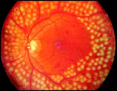
Step 3: Laser Application
The core step in PRP involves the precise application of laser therapy to the peripheral retina. Using a specialized laser system, the ophthalmologist delivers numerous small burns to targeted areas of the retina, typically in a scattered pattern. This laser treatment aims to reduce abnormal blood vessel growth and promote retinal vascular stability, thus preventing further complications.
Step 4: Monitoring
Throughout the PRP procedure, continuous monitoring of the patient’s response is essential. The ophthalmologist evaluates the effectiveness of laser therapy, adjusts treatment parameters as needed, and ensures patient safety and comfort. Real-time feedback allows for modifications to optimize treatment outcomes and minimize potential complications.
Step 5: Post-Procedure Care
Following PRP, appropriate post-procedure care is crucial to promote healing and minimize complications. Patients may experience mild discomfort, blurred vision, or sensitivity to light immediately after PRP. Eye drops may be prescribed to reduce inflammation and prevent infection. Patients should be advised on post-procedure instructions, including the use of prescribed medications and follow-up appointments.
Lasers Used:
PRP traditionally utilizes argon or diode lasers for treatment. Alternative therapies may also employ laser technology but with variations in wavelength, intensity, or delivery methods to optimize treatment efficacy and patient comfort.
Complications of PRP:
While panretinal photocoagulation (PRP) is an effective treatment for retinal disorders, it is not without potential complications. Understanding these complications is essential for ophthalmologists to mitigate risks and optimize patient outcomes. Let’s explore the common complications associated with PRP and their implications for clinical practice.
Reduced Night Vision:
One potential complication of PRP is reduced night vision, particularly in patients with extensive laser treatment. The creation of laser scars in the peripheral retina may lead to a reduction in peripheral vision, impacting night vision and causing difficulty seeing in dimly lit environments. Patients should be counseled about this potential side effect, and careful consideration should be given to treatment parameters to minimize its occurrence.
Retinal Scarring:
Excessive laser therapy during PRP can lead to retinal scarring, which may affect visual function and overall retinal health. While the goal of PRP is to induce controlled scar formation to stabilize the retina and reduce neovascularization, overzealous treatment can result in extensive scarring, compromising retinal integrity. Ophthalmologists must carefully titrate laser intensity and duration to achieve therapeutic goals while minimizing the risk of excessive scarring.
Macular Edema:
In some cases, PRP may induce macular edema, particularly in patients with pre-existing diabetic macular edema or compromised blood-retinal barriers. The inflammatory response triggered by laser therapy can lead to fluid accumulation in the macula, causing temporary or permanent vision loss. Close monitoring of patients following PRP is essential to detect and manage macular edema promptly, utilizing interventions such as anti-inflammatory medications or intravitreal injections as needed.
Infection:
Although rare, infection is a potential complication of PRP, particularly if proper aseptic techniques are not followed during the procedure. Corneal epithelial defects or conjunctival inflammation can increase the risk of microbial contamination, leading to endophthalmitis or other ocular infections. Ophthalmologists should adhere to strict sterile protocols during PRP and promptly address any signs or symptoms of infection in treated patients.
Alternative Treatments:
- Anti-VEGF Therapy: Intravitreal injections of anti-vascular endothelial growth factor (VEGF) medications have revolutionized the management of retinal diseases. These injections target abnormal blood vessel growth, reducing leakage and edema without the need for laser therapy. They are particularly effective in diabetic macular edema and wet age-related macular degeneration.
- Steroid Implants: Intravitreal steroid implants offer sustained release of corticosteroids, providing anti-inflammatory and anti-angiogenic effects. They are indicated for conditions such as diabetic macular edema and uveitis, offering an alternative to laser therapy with fewer treatment sessions and less risk of retinal scarring.
- Vitreoretinal Surgery: In cases of severe retinal detachment or tractional retinal disorders, vitreoretinal surgery may be necessary to repair the retina. Surgical techniques such as vitrectomy and membrane peeling can address underlying pathology and restore retinal anatomy, reducing the need for extensive laser treatment.
Follow-Up
Regardless of the treatment modality chosen, regular follow-up is essential to monitor disease progression, assess treatment response, and address any complications that may arise. Close collaboration between patients and healthcare providers ensures optimal outcomes in the management of retinal disorders.
In summary, while PRP remains a cornerstone in the treatment of retinal diseases, alternative modalities offer valuable options for patients and physicians. By understanding the indications, procedural steps, potential complications, and alternative treatments available, healthcare providers can tailor therapy to individual patient needs, ultimately optimizing visual outcomes and preserving retinal function.


Pingback: Home - ECG Oxford