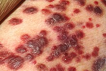
Kaposi sarcoma;
Quick Facts :
- Kaposi Sarcoma (KS) is a rare cancer caused by human herpesvirus 8 (HHV-8).
- It manifests as purplish skin lesions and can affect internal organs.
- Four types exist: classic, endemic African, HIV-related, and iatrogenic.
- Diagnosis involves biopsy and HHV-8 serology.
- Treatment includes local therapies, chemotherapy, and HAART for HIV-related KS.

Overview
Kaposi Sarcoma (KS) stands as a notable malignancy, characterized by its multifaceted nature and distinct presentation.
What is Kaposi Sarcoma?
Kaposi Sarcoma manifests as a rare form of cancer, primarily affecting the skin, mucous membranes, and sometimes internal organs. It emerges from the abnormal growth of cells, particularly the endothelial cells lining blood vessels and lymphatic vessels.
Types of Kaposi Sarcoma:
Kaposi Sarcoma (KS) exhibits a multifaceted spectrum, comprising four distinct clinical forms, each characterized by unique features and natural histories.
1. Classic Form
- Affected Population: Typically observed in elderly men hailing from Mediterranean and Eastern European backgrounds.
- Primary Location: Predominantly manifests on the lower extremities.
- Clinical Presentation: Presents as solitary or multiple skin lesions, often in the form of purplish or reddish-brown nodules.
- Natural History: Tends to follow an indolent course, with a slow progression and relatively favorable prognosis.
2. Endemic African Form
- Affected Population: Primarily affects children in endemic regions, particularly in sub-Saharan Africa.
- Characteristics: Characterized by generalized lymph node involvement and systemic manifestations.
- Presentation: Lesions may appear on the skin, mucous membranes, and internal organs.
- Natural History: Varied, with some cases exhibiting indolent progression while others manifesting a more aggressive course.
3. HIV-Related Form
- Affected Population: Occurs predominantly in individuals living with HIV/AIDS, particularly those not receiving highly active antiretroviral therapy (HAART).
- Extent of Involvement: Presents with diffuse involvement of the skin and internal organs.
- Clinical Features: Lesions may be widespread, affecting multiple areas of the body including the skin, mucous membranes, and visceral organs.
- Natural History: Exhibits a diverse spectrum, ranging from indolent to aggressive and potentially fatal, particularly in anaplastic varieties.
4. Iatrogenic Form
- Affected Population: Arises in immunosuppressed individuals, often following organ transplantation or prolonged immunosuppressive therapy.
- Similarities with HIV-Related Form: Shares similarities with the HIV-related form, including diffuse skin and visceral involvement.
- Risk Factors: Associated with the use of immunosuppressive medications.
- Natural History: Varied, with some cases demonstrating a relatively indolent course, while others may progress rapidly, necessitating aggressive intervention.
Causes of Kaposi Sarcoma
The human herpesvirus 8 (HHV-8), also known as Kaposi’s sarcoma-associated herpesvirus (KSHV), serves as the primary culprit behind the development of Kaposi Sarcoma. This virus infects cells, prompting uncontrolled cellular proliferation and the formation of characteristic lesions.
Etiopathogenesis of Kaposi Sarcoma:
HHV-8: The Viral Culprit
- Viral Structure: Double-stranded enveloped DNA virus with six major subtypes (A-F).
- Transmission: Primarily via saliva in childhood and sexually, with occasional cases via blood transfusion or intravenous drug use.
- Co-evolution with Human Population: Requires immune defects and inflammation for malignancy development.
Viral Pathogenesis
- Endothelial Cell Infection: HHV-8 infects endothelial cells, activating the mTOR pathway.
- Mesenchymal Differentiation: Alters cells to have mesenchymal differentiation.
- Aberrant Angiogenesis: Promotes aberrant angiogenesis, contributing to lesion formation.
Immune Suppression and Inflammation
- Persistence and Proliferation: HHV-8 infected cells persist and proliferate due to immune suppression and inflammation.
- Role of Latency-associated Nuclear Antigen (LANA): LANA inhibits apoptosis by binding p53 and maintains the viral episome.
Molecular Mechanisms
- NF-kB Activation: HHV-8 activates NF-kB, upregulating cytokine expression, including VEGF and bFGF.
- Neo-angiogenesis: Upregulation of VEGF, bFGF induces neo-angiogenesis.
- Spindled Morphology: Induces c-kit expression and produces the spindled morphology of Kaposi sarcoma cells.
- Matrix Metalloproteinases: HHV-8 upregulates matrix metalloproteinases, contributing to lesion progression.
Clinical Presentation
- Vascular Lesions: Presents as violaceous pink to purple plaques on the skin or mucocutaneous surfaces.
- Stages of Lesions: Progresses through patch, plaque, and nodule stages.
- Associated Symptoms: May be painful with associated lymphedema and secondary infection.
Systemic Involvement
- Extrapulmonary Manifestations: Besides lymph nodes, Kaposi sarcoma affects the lungs, gastrointestinal system, and other visceral organs.
- Respiratory Complications: Respiratory involvement can be associated with death due to Kaposi sarcoma.
Risk Factors:
- Immunocompromised States: Individuals with weakened immune systems, such as those living with HIV/AIDS or undergoing immunosuppressive therapy post-organ transplantation, face heightened susceptibility.
- Geographic Prevalence: Geographical regions with higher rates of HHV-8 transmission, such as sub-Saharan Africa, exhibit increased incidence rates.
Signs and Symptoms of Kaposi Sarcoma
- Skin Lesions: The most visible indication, appearing as purplish or reddish-brown nodules or patches on the skin, mouth, or genitals.
- Mucosal Involvement: Lesions may also manifest internally, affecting the mucous membranes of the mouth, throat, or gastrointestinal tract.
- Systemic Symptoms: In advanced stages, patients may experience symptoms such as fever, weight loss, and fatigue.
Diagnosis :
Clinical Evaluation
- Visual Inspection: Examination of the skin and mucous membranes for characteristic lesions, which may present as purplish or reddish-brown nodules or patches.
- Assessment of Symptoms: Evaluation of associated symptoms such as pain, lymphedema, or secondary infection.
Diagnostic Procedures
- Biopsy: Definitive diagnosis often requires a tissue biopsy, where a small sample of the lesion is obtained and examined under a microscope.
- Types of Biopsy: Excisional biopsy, incisional biopsy, or punch biopsy may be performed based on the size and location of the lesion.
- Imaging Studies: Imaging modalities such as ultrasound, computed tomography (CT), or magnetic resonance imaging (MRI) may be utilized to assess the extent of disease involvement, particularly in cases with internal organ manifestations.
Laboratory Tests
- HHV-8 Serology: Testing for antibodies to human herpesvirus 8 (HHV-8) can aid in confirming the diagnosis, particularly in HIV-related Kaposi Sarcoma.
- HIV Testing: Given the association between HIV infection and Kaposi Sarcoma, HIV testing is routinely performed in suspected cases.
Differential Diagnosis
- Other Vascular Lesions: Lesions such as hemangiomas, angiosarcomas, or bacillary angiomatosis may resemble Kaposi Sarcoma and require differentiation.
- Infectious Etiologies: Conditions such as cutaneous tuberculosis, histoplasmosis, or syphilis may mimic Kaposi Sarcoma clinically and histologically.
Staging
- TNM Classification: Staging of KS follows the TNM classification system, assessing the extent of tumor (T), lymph node involvement (N), and metastasis (M).
- Clinical Staging: Integration of clinical and radiological findings to determine the stage of disease and guide treatment decisions.
Treatment:
Localized Skin Involvement
- Local Excision: Surgical removal of affected skin lesions.
- Liquid Nitrogen: Cryotherapy using liquid nitrogen to freeze and destroy lesions.
- Vincristine Injection: Direct injection of vincristine into lesions to target cancerous cells.
Chemotherapy for Endemic and Systemic Forms
- Mainstay of Treatment: Chemotherapy serves as a primary treatment modality, particularly for endemic and systemic forms, especially in children.
- Agents Used: Liposomal anthracyclines, paclitaxel, etoposide, vincristine, vinblastine, vinorelbine, bleomycin, or combinations such as doxorubicin, bleomycin, and vincristine.
- Interferon-alpha: Interferon-alpha 2b has shown efficacy in some cases.
HIV-Related Kaposi Sarcoma
- HAART Therapy: High-activity antiretroviral therapy (HAART) is pivotal in managing HIV-related KS.
- Regression of Tumors: HAART treatment can lead to regression or complete treatment of KS lesions.
Iatrogenic Kaposi Sarcoma
- Balancing Immune Suppression: Treatment involves balancing reduction in immune suppression or withdrawal of steroid treatment with the risk of transplant rejection and sarcoma treatment.
Classical Kaposi Sarcoma
- Radiation Therapy: Radiotherapy is effective, especially in early stages.
- Recommended Dosages: Vary based on the location of lesions, ranging from 15.2 Gy for oral lesions to 30 Gy with hypofractionation for other body parts.
- Palliative Care: Radiation may be used palliatively in later stages to reduce pain, edema, and bleeding.
Medical Oncology Approach:
- Local Therapies: Sclerotherapy, intralesional vinca-alkaloids, bleomycin, interferon-alpha, topical alitretinoin, or imiquimod cream may be used for localized disease.
- Systemic Chemotherapy: Liposomal anthracyclines, paclitaxel, etoposide, vincristine, vinblastine, vinorelbine, bleomycin, or combination therapies are employed for systemic disease.
- Other Investigational Therapies: Thalidomide, VEGF inhibitors, tyrosine kinase inhibitors, and matrix metalloproteinase inhibitors are under investigation as potential treatment options.
Prognosis :
The prognosis of KS varies significantly depending on various factors such as the extent of disease, immune status, and response to treatment. With early diagnosis and appropriate management, many individuals can achieve remission and maintain a good quality of life.
In summary, Kaposi Sarcoma remains a complex entity, intertwined with viral etiology, immune status, and clinical presentation.
Read more:

