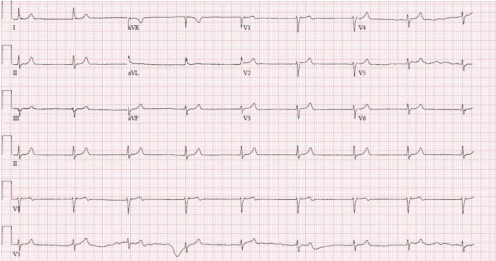
A 35-year-old woman presented to the emergency department with a sudden onset of fatigue, bruising, and confusion. She reported a history of easy bruising over the past week, accompanied by fever and abdominal pain.
Examination Findings
During the physical examination, the following observations were made:
- General Appearance: The patient appeared pale and fatigued.
- Vital Signs: Blood pressure was elevated at 150/90 mmHg, pulse rate was 110 beats per minute, and she had a fever of 38.5°C.
- Skin: Multiple petechiae and ecchymoses were noted on her limbs and trunk.
- Neurological: The patient was confused and exhibited mild hemiparesis on the right side.
- Abdomen: Mild tenderness was present without any palpable organomegaly.
Laboratory Results
The initial laboratory tests revealed:
- Complete Blood Count (CBC):
- Platelet count: 20,000/µL (normal: 150,000-450,000/µL)
- Hemoglobin: 8.0 g/dL (normal: 12-16 g/dL for women)
- White blood cells (WBC): 10,000/µL (normal: 4,000-11,000/µL)
- Peripheral Blood Smear: Presence of schistocytes (fragmented red blood cells).

- Renal Function:
- Creatinine: 2.5 mg/dL (normal: 0.6-1.2 mg/dL)
- Blood urea nitrogen (BUN): 35 mg/dL (normal: 7-20 mg/dL)
- Coagulation Profile:
- Prothrombin time (PT): 12 seconds (normal: 11-13.5 seconds)
- Partial thromboplastin time (PTT): 30 seconds (normal: 25-35 seconds)
Additional Laboratory Tests
- Lactate Dehydrogenase (LDH): 1,500 U/L (normal: 140-280 U/L), significantly elevated, consistent with hemolysis.
- Haptoglobin: < 10 mg/dL (normal: 30-200 mg/dL), indicating hemolytic anemia.
- ADAMTS13 Activity: < 5% (normal: > 67%), severely reduced, with autoantibodies against ADAMTS13 present.
Radiology
While radiology is not always central in diagnosing TTP, certain imaging studies can aid in evaluating organ damage:
- CT Scan of the Head: Showed no acute intracranial hemorrhage but indicated mild cerebral edema, correlating with the patient’s confusion and hemiparesis.
- Ultrasound of the Abdomen: Revealed no significant abnormalities, ruling out other causes of abdominal pain.
Diagnosis
The diagnosis of TTP is primarily clinical, supported by laboratory findings and reduced ADAMTS13 activity. Key criteria for TTP include:
- Microangiopathic Hemolytic Anemia: Evidence of hemolysis (schistocytes on smear, elevated LDH).
- Thrombocytopenia: Low platelet count without another apparent cause.
- Organ Dysfunction: Neurological symptoms, renal impairment, and fever are common presentations.
Differential Diagnosis
While evaluating TTP, other conditions to consider include:
- Hemolytic Uremic Syndrome (HUS): Typically seen in children, associated with E. coli infections.
- Disseminated Intravascular Coagulation (DIC): Characterized by widespread clotting and bleeding, usually secondary to another condition like sepsis.
- Systemic Lupus Erythematosus (SLE): Autoimmune disease that can present with similar hematologic abnormalities.
Treatment and Management
Immediate treatment is crucial for TTP due to its rapid progression and high mortality if untreated:
- Plasma Exchange (PEX): The mainstay of treatment, replacing the patient’s plasma to remove autoantibodies and provide functional ADAMTS13.
- Corticosteroids: To suppress the immune response.
- Rituximab: For refractory cases, targeting B cells producing the autoantibodies.
- Supportive Care: Including red blood cell transfusions and dialysis if renal failure occurs.
Conclusion
TTP is a medical emergency requiring prompt recognition and treatment. With advancements in understanding and managing TTP, the prognosis has significantly improved. However, early diagnosis and intervention remain key to reducing morbidity and mortality associated with this condition.
