Cerebellar signs;
Introduction:
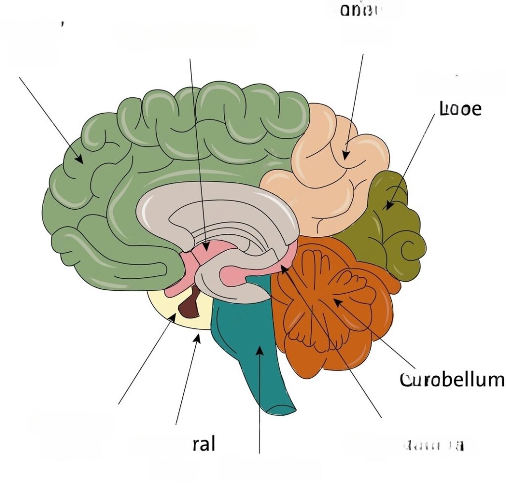
The cerebellum, nestled in the posterior cranial fossa, serves as a pivotal hub for coordinating movements, intricately connected to the brainstem, basal ganglia, and cerebral cortex. Lesions within this intricate structure give rise to discernible neurological signs, shedding light on underlying pathology and guiding diagnostic and management strategies.
Common Cerebellar Neurological Signs
- Extraocular Movements: Testing Eye Movements
- Nystagmus: Central nystagmus differs from its peripheral counterpart, often exhibiting bidirectional patterns and exacerbated responses with gaze changes. Evaluation involves observing the pattern and direction of nystagmus, which worsens when gaze is directed towards the affected side (Alexander law). Causes include lesions affecting the vestibulocerebellar pathways.
- Impaired Smooth Pursuits: Cerebellar lesions disrupt smooth eye tracking, leading to catch-up saccades instead. Findings include inability to track moving objects smoothly. Lesions in the cerebellar ocular motor tracts contribute to this impairment.
- HINTS Exam: A combination of Head Impulse test, Nystagmus assessment, and Test of Skew aids in distinguishing central from peripheral etiologies of vertigo. This includes observing for corrective saccades in response to head movements, bi-directional nystagmus, and vertical malalignment of the eyes (skew deviation). Central causes involve lesions within the cerebellum or brainstem.
- Motor Coordination:
- Dysmetria: Manifestations include overshooting or undershooting targets, evident in tests like the Finger-to-Nose and Heel-to-Shin tests. Testing involves assessing the accuracy of reaching movements. Cerebellar hemisphere lesions disrupt the coordination necessary for precise movements.
- Adiadochokinesia: Impaired execution of rapid alternating movements, observed in patients with cerebellar lesions. The examiner asks the patient to perform rapid pronation and supination of the forearm. Jerky and irregular movements indicate cerebellar dysfunction, often due to lesions affecting the cerebellar motor pathways.
- Rebound Phenomenon: Exaggerated flexion of joints upon sudden cessation of resistance, indicative of cerebellar dysfunction. This is tested by resisting flexion of joints and then suddenly releasing the resistance. Failure of timely contraction of antagonist muscles results in exaggerated flexion, often seen in cerebellar lesions.
- Intention Tremor:
- Intention Tremor: A kinetic tremor exacerbated during purposeful movements, notably observed in the Finger-to-Nose test. Findings include tremor worsening as the target is approached. Cerebellar lesions disrupt the fine motor control required for precise movements.
- Ambulation:
- Stance and Posture: Patients often exhibit a broad-based stance and titubation, characterized by swaying while standing. This is observed during the physical examination. Cerebellar lesions affect the coordination necessary for maintaining balance.
- Gait: Ataxic gait, resembling acute alcohol intoxication, manifests as staggering or swaying during locomotion. Differential diagnoses include various conditions affecting the cerebellum, such as hydrocephalus, and infectious etiologies. Observation of gait characteristics aids in identifying cerebellar dysfunction.
- Tandem Walk: Inability to walk in a straight line with a heel-to-toe pattern, indicative of cerebellar dysfunction. The examiner asks the patient to walk in a straight line with the heel of the leading foot touching the toes of the lagging foot. Cerebellar lesions disrupt the coordination necessary for tandem walking.
- Romberg’s Sign: Absence of disproportionate swaying with eyes closed compared to open, indicative of cerebellar lesions. This is assessed by asking the patient to stand with feet together and eyes closed. Excessive swaying, even with eyes open, can be seen in cerebellar lesions due to impaired balance regulation.
- Hypotonia:
- Ipsilateral Hypotonia: Damage to half of the cerebellum results in reduced muscle tone on the same side. This is observed during the physical examination and results from disruption of cerebellar pathways involved in regulating muscle tone.
- Cerebellar Mutism:
- Central Cerebellar Injury: Lesions within the central cerebellum may lead to temporary or persistent mutism, impacting communication. This is observed following injuries or surgeries involving the central cerebellum, disrupting speech production pathways.
Summary:
In summary ,cerebellar neurological signs serve as invaluable markers of cerebellar dysfunction, offering crucial insights into motor coordination and balance regulation.
Read more: